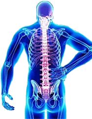
Back and Spine Anatomy
-
- Movement
- Balance
- Upright posture
- Spinal cord protection
- Shock absorption
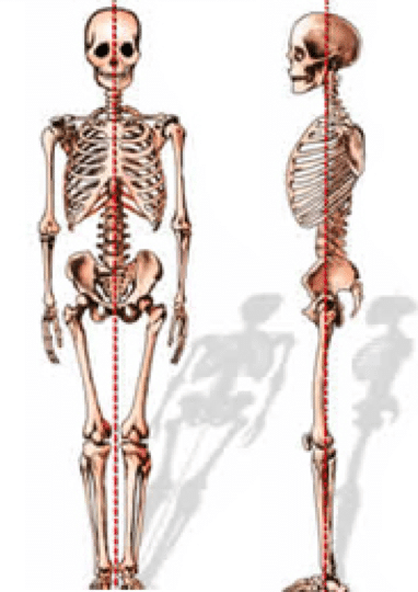
The normal adult spine is balanced over the pelvis, requiring minimal workload on the muscles to maintain an upright posture.
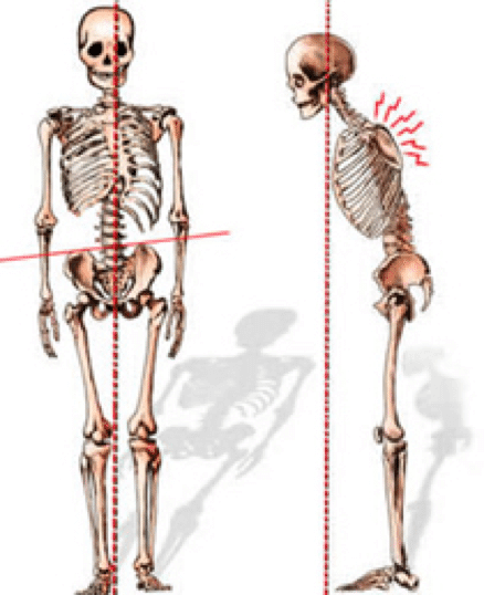
Loss of spinal balance can result in strain to the spinal muscles and spinal deformity. When the spine is injured and its function impaired, the consequences may be painful and even disabling.
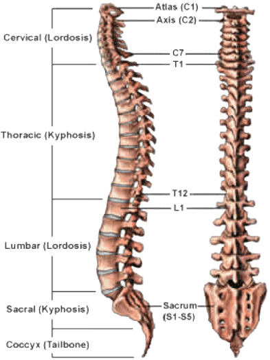
Regions of the Spine
Humans are born with 33 separate vertebrae. By adulthood, we typically have 24 due to the fusion of the vertebrae in the sacrum.
-
- The top 7 vertebrae that form the neck are called the cervical spine and are labeled C1-C7. The seven vertebrae of the cervical spine are responsible for the normal function and mobility of the neck. They also protect the spinal cord, nerves and arteries that extend from the brain to the rest of the body.
- The upper back, or thoracic spine, has 12 vertebrae, labeled T1-T12.
- The lower back, or lumbar spine, has 5 vertebrae, labeled L1-L5. The lumbar spine bears the most weight relative to other regions of the spine, which makes it a common source of back pain.
- The sacrum (S1) and coccyx (tailbone) are made up of 9 vertebrae that are fused together to form a solid, bony unit.
Spinal Curvature
When viewed from the front or back, the normal spine is in a straight line, with each vertebra sitting directly on top of the other. Curvature to one side or the other indicates a condition called scoliosis.
When viewed from the side, the normal spine has three gradual curves:
-
- The neck has a lordotic curve, meaning that it curves inward.
- The thoracic spine has a kyphotic curve, meaning it curves outward.
- The lumbar spine also has a lordotic curve.
These curves help the spine to support the load of the head and upper body, and maintain balance in the upright position. Excessive curvature, however, may result in spinal imbalance.
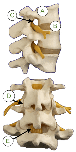
Elements of the Spine
The elements of the spine are designed to protect the spinal cord, support the body and facilitate movement.
The spinal cord is covered by a protective membrane called the dura mater, which forms a watertight sac around the spinal cord and nerves. Inside this sac is spinal fluid, which surrounds the spinal cord.
The nerves in each area of the spinal cord are connected to specific parts of the body. Those in the cervical spine, for example, extend to the upper chest and arms; those in the lumbar spine the hips, buttocks and legs. The nerves also carry electrical signals back to the brain, creating sensations. Damage to the nerves, nerve roots or spinal cord may result in symptoms such as pain, tingling, numbness and weakness, both in and around the damaged area and in the extremities.
Spinal Muscles
Many muscle groups that move the trunk and the limbs also attach to the spinal column. The muscles that closely surround the bones of the spine are important for maintaining posture and helping the spine to carry the loads created during normal activity, work and play. Strengthening these muscles can be an important part of physical therapy and rehabilitation.
Nervous System
All of the elements of the spinal column and vertebrae serve the purpose of protecting the spinal cord, which provides communication to the brain, mobility and sensation in the body through the complex interaction of bones, ligaments and muscle structures of the back and the nerves that surround it.
The true spinal cord ends at approximately the L1 level, where it divides into the many different nerve roots that travel to the lower body and legs. This collection of nerve roots is called the cauda equina, which means “horse’s tail,” and describes the continuation of the nerve roots at the end of the spinal cord.
Don’t Suffer Any Longer…